Magnetic Resonance Imaging Unit
Magnetic Resonance Imaging (MRI) uses magnetic fields and radio waves to create images of bodies or other items placed in the bore of the MRI scanner. There are a number of different imaging modalities supported by MRI and which we use at CINN.
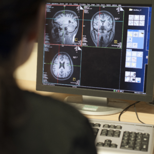
These modalities include structural MRI, in which high-resolution images can be used, for example, to describe the health status of the grey and white matter of the brain and other features of its morphology, such as the ventricles. In contrast to structural MRI, functional MRI (fMRI) measures changes in brain activity over time, or in response to an external stimulus or an internally generated action, by measuring the levels of oxygen in specific parts of the brain. The amount of oxygen a brain area consumes is proportional to the energy it uses, offering a window into the dynamics of regional brain function.
We also use other MRI techniques: for example, Arterial Spin Labeling (ASL) provides information about the flow of blood to and from different brain regions; diffusion MRI (dMRI) uses Diffusion Tensor Imaging (DTI) to provide a detailed picture of the structural connections between brain regions. We also employ Magnetic Resonance Spectroscopy (MRS), which measures the chemical composition of brain tissue. Functional MRS (fMRS) extends this approach by enabling the assessment of metabolite changes over time or in response to stimuli.
CINN houses a state-of-the-art Siemens MAGNETOM Prisma-fit 3T MRI Scanner, with a large selection of coils for clinical and research use. A dedicated stimulus presentation PC with MRI-compatible stimulus response pads and joysticks can be used to measure participant responses to behavioural and cognitive tasks presented on the screen. Supplementary instruments measuring physiological features, including eye tracking, skin conductance, breathing rate/volume, allow for real-time data performance monitoring and for the co-measurement of physiology and brain function.
Our new functional near infrared spectroscopy (fNIRS) systems measure oxygenated and deoxygenated blood flow across the surface of the cortex using near infrared light. This signal is equivalent to the BOLD response measured using fMRI, reflecting brain metabolism as a proxy for brain activity. The system we own, the Artinis MediBrite C system, is the only medically-certified fNIRS system available, making it ideal for clinical usage. The equipment is portable, wireless, movable, and lightweight, and so can be used anywhere, allowing for more ecologically valid and naturalistic data collection. It is also suitable for adults, children, and infants, making it an ideal neuroimaging system regardless of the age of participants.
We own two Artinis MediBrite C systems, allowing us to conduct fNIRS hyperscanning – simultaneous two-person data acquisition). The caps are also compatible with Brain Products ActiCAP snap EEG electrodes, meaning that we can also acquire simultaneous fNIRS and EEG data, and with neurostimulation equipment such as transcranial magnetic stimulation (TMS) and transcranial direct current stimulation (TDCS) for causal inference. The MediBrite system can record up to 27 channels at once, and has a battery and internal storage to last for 2 hours of continuous recording. We also have licenses for Artinis’ Brite Connect recording software, and OxySoft 4 recording and analysis software, so we can support fNIRS studies end-to-end, from scoping and development to processing and analysis.